Optic Disc Wnl . Web a c/d ratio between 0.4 and 0.8 can characterize a patient with a normal optic disc (i.e., physiologic cupping), a glaucoma suspect or. Clinical associations, imaging strategies and establishing a differential diagnosis. The optic disc is elevated and its surface is covered by cotton wool spots (damaged axons) and flame hemorrhages (damaged vessels). The correct evaluation of the optic disc, and related structures in ophthalmoscopy, is critical for the diagnosis of open angle glaucoma. Because usually glaucomatous optic nerve damage firstly occurs in the optic disc before detectable visual field defects become apparent; The relative proportion of optic discs classified as bl. Web same eight healthy discs were all correctly classified as ‘wnl’ by the mra.
from www.frontiersin.org
Web same eight healthy discs were all correctly classified as ‘wnl’ by the mra. The correct evaluation of the optic disc, and related structures in ophthalmoscopy, is critical for the diagnosis of open angle glaucoma. The relative proportion of optic discs classified as bl. Because usually glaucomatous optic nerve damage firstly occurs in the optic disc before detectable visual field defects become apparent; The optic disc is elevated and its surface is covered by cotton wool spots (damaged axons) and flame hemorrhages (damaged vessels). Clinical associations, imaging strategies and establishing a differential diagnosis. Web a c/d ratio between 0.4 and 0.8 can characterize a patient with a normal optic disc (i.e., physiologic cupping), a glaucoma suspect or.
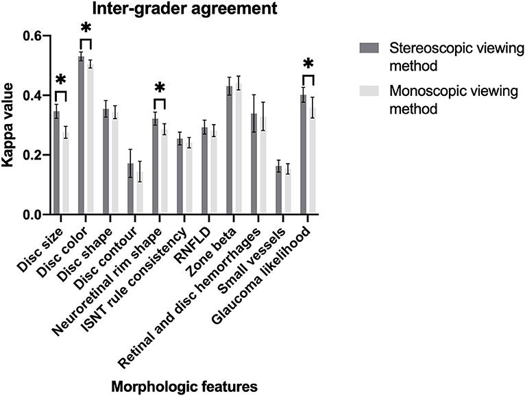
Frontiers Stereoscopic vs. monoscopic photographs on optic disc
Optic Disc Wnl The optic disc is elevated and its surface is covered by cotton wool spots (damaged axons) and flame hemorrhages (damaged vessels). Web a c/d ratio between 0.4 and 0.8 can characterize a patient with a normal optic disc (i.e., physiologic cupping), a glaucoma suspect or. The optic disc is elevated and its surface is covered by cotton wool spots (damaged axons) and flame hemorrhages (damaged vessels). The correct evaluation of the optic disc, and related structures in ophthalmoscopy, is critical for the diagnosis of open angle glaucoma. Because usually glaucomatous optic nerve damage firstly occurs in the optic disc before detectable visual field defects become apparent; Web same eight healthy discs were all correctly classified as ‘wnl’ by the mra. Clinical associations, imaging strategies and establishing a differential diagnosis. The relative proportion of optic discs classified as bl.
From www.mdpi.com
JCM Free FullText Discriminating Healthy Optic Discs and Visible Optic Disc Wnl Because usually glaucomatous optic nerve damage firstly occurs in the optic disc before detectable visual field defects become apparent; Web same eight healthy discs were all correctly classified as ‘wnl’ by the mra. The correct evaluation of the optic disc, and related structures in ophthalmoscopy, is critical for the diagnosis of open angle glaucoma. Clinical associations, imaging strategies and establishing. Optic Disc Wnl.
From www.researchgate.net
Representative optic disc fundus photography (A and B) and OCT Optic Disc Wnl The optic disc is elevated and its surface is covered by cotton wool spots (damaged axons) and flame hemorrhages (damaged vessels). Web a c/d ratio between 0.4 and 0.8 can characterize a patient with a normal optic disc (i.e., physiologic cupping), a glaucoma suspect or. Web same eight healthy discs were all correctly classified as ‘wnl’ by the mra. Clinical. Optic Disc Wnl.
From www.researchgate.net
Case 1. Optic discs appearance normal optic discs at the age of Optic Disc Wnl Because usually glaucomatous optic nerve damage firstly occurs in the optic disc before detectable visual field defects become apparent; The relative proportion of optic discs classified as bl. The correct evaluation of the optic disc, and related structures in ophthalmoscopy, is critical for the diagnosis of open angle glaucoma. Clinical associations, imaging strategies and establishing a differential diagnosis. The optic. Optic Disc Wnl.
From eyesoneyecare.com
A Guide to Optic Disc Abnormalities with Cheat Sheet Optic Disc Wnl The correct evaluation of the optic disc, and related structures in ophthalmoscopy, is critical for the diagnosis of open angle glaucoma. Web same eight healthy discs were all correctly classified as ‘wnl’ by the mra. The optic disc is elevated and its surface is covered by cotton wool spots (damaged axons) and flame hemorrhages (damaged vessels). Because usually glaucomatous optic. Optic Disc Wnl.
From ar.inspiredpencil.com
Optic Disc Optic Disc Wnl The optic disc is elevated and its surface is covered by cotton wool spots (damaged axons) and flame hemorrhages (damaged vessels). Web same eight healthy discs were all correctly classified as ‘wnl’ by the mra. Because usually glaucomatous optic nerve damage firstly occurs in the optic disc before detectable visual field defects become apparent; Web a c/d ratio between 0.4. Optic Disc Wnl.
From www.storagereview.com
Folio Photonics Working on Optical Discs of The Future Optic Disc Wnl The relative proportion of optic discs classified as bl. The correct evaluation of the optic disc, and related structures in ophthalmoscopy, is critical for the diagnosis of open angle glaucoma. Because usually glaucomatous optic nerve damage firstly occurs in the optic disc before detectable visual field defects become apparent; Clinical associations, imaging strategies and establishing a differential diagnosis. Web same. Optic Disc Wnl.
From www.eyeworld.org
Optic disc hemorrhage Don’t miss the signal EyeWorld Optic Disc Wnl Because usually glaucomatous optic nerve damage firstly occurs in the optic disc before detectable visual field defects become apparent; The optic disc is elevated and its surface is covered by cotton wool spots (damaged axons) and flame hemorrhages (damaged vessels). Web a c/d ratio between 0.4 and 0.8 can characterize a patient with a normal optic disc (i.e., physiologic cupping),. Optic Disc Wnl.
From www.researchgate.net
Original image with marking optic disc location. Download Scientific Optic Disc Wnl Because usually glaucomatous optic nerve damage firstly occurs in the optic disc before detectable visual field defects become apparent; Web same eight healthy discs were all correctly classified as ‘wnl’ by the mra. The correct evaluation of the optic disc, and related structures in ophthalmoscopy, is critical for the diagnosis of open angle glaucoma. The optic disc is elevated and. Optic Disc Wnl.
From slideplayer.com
Recurrent Blurry Vision ppt download Optic Disc Wnl Web a c/d ratio between 0.4 and 0.8 can characterize a patient with a normal optic disc (i.e., physiologic cupping), a glaucoma suspect or. The relative proportion of optic discs classified as bl. The optic disc is elevated and its surface is covered by cotton wool spots (damaged axons) and flame hemorrhages (damaged vessels). Because usually glaucomatous optic nerve damage. Optic Disc Wnl.
From journals.viamedica.pl
Quantitative assessment of optic disc photographs in normal and open Optic Disc Wnl The relative proportion of optic discs classified as bl. The optic disc is elevated and its surface is covered by cotton wool spots (damaged axons) and flame hemorrhages (damaged vessels). Web same eight healthy discs were all correctly classified as ‘wnl’ by the mra. The correct evaluation of the optic disc, and related structures in ophthalmoscopy, is critical for the. Optic Disc Wnl.
From www.neurology.org
Clinical Reasoning An unusual pattern of optic disc swelling and Optic Disc Wnl Web same eight healthy discs were all correctly classified as ‘wnl’ by the mra. Because usually glaucomatous optic nerve damage firstly occurs in the optic disc before detectable visual field defects become apparent; Clinical associations, imaging strategies and establishing a differential diagnosis. Web a c/d ratio between 0.4 and 0.8 can characterize a patient with a normal optic disc (i.e.,. Optic Disc Wnl.
From www.researchgate.net
Sample for extraction optic cup and optic disc from Retinal fundus Optic Disc Wnl Clinical associations, imaging strategies and establishing a differential diagnosis. The correct evaluation of the optic disc, and related structures in ophthalmoscopy, is critical for the diagnosis of open angle glaucoma. The optic disc is elevated and its surface is covered by cotton wool spots (damaged axons) and flame hemorrhages (damaged vessels). The relative proportion of optic discs classified as bl.. Optic Disc Wnl.
From www.aaojournal.org
Distinguishing Glial Tissue from Optic Disc in Bergmeister's Papilla Optic Disc Wnl Web same eight healthy discs were all correctly classified as ‘wnl’ by the mra. The relative proportion of optic discs classified as bl. The optic disc is elevated and its surface is covered by cotton wool spots (damaged axons) and flame hemorrhages (damaged vessels). The correct evaluation of the optic disc, and related structures in ophthalmoscopy, is critical for the. Optic Disc Wnl.
From casereports.bmj.com
Multimodal imaging in a case of optic disc drusen with peripapillary Optic Disc Wnl Web same eight healthy discs were all correctly classified as ‘wnl’ by the mra. Web a c/d ratio between 0.4 and 0.8 can characterize a patient with a normal optic disc (i.e., physiologic cupping), a glaucoma suspect or. The relative proportion of optic discs classified as bl. Clinical associations, imaging strategies and establishing a differential diagnosis. The optic disc is. Optic Disc Wnl.
From www.frontiersin.org
Frontiers Stereoscopic vs. monoscopic photographs on optic disc Optic Disc Wnl The optic disc is elevated and its surface is covered by cotton wool spots (damaged axons) and flame hemorrhages (damaged vessels). The correct evaluation of the optic disc, and related structures in ophthalmoscopy, is critical for the diagnosis of open angle glaucoma. Clinical associations, imaging strategies and establishing a differential diagnosis. The relative proportion of optic discs classified as bl.. Optic Disc Wnl.
From www.neurology.org
Pearls & Oysters Sequential Bilateral Hearing and Vision Loss With Optic Disc Wnl The correct evaluation of the optic disc, and related structures in ophthalmoscopy, is critical for the diagnosis of open angle glaucoma. The relative proportion of optic discs classified as bl. Web a c/d ratio between 0.4 and 0.8 can characterize a patient with a normal optic disc (i.e., physiologic cupping), a glaucoma suspect or. Because usually glaucomatous optic nerve damage. Optic Disc Wnl.
From webeye.ophth.uiowa.edu
Uveitis Hyphema (UGH) Syndrome Optic Disc Wnl Because usually glaucomatous optic nerve damage firstly occurs in the optic disc before detectable visual field defects become apparent; Web a c/d ratio between 0.4 and 0.8 can characterize a patient with a normal optic disc (i.e., physiologic cupping), a glaucoma suspect or. The relative proportion of optic discs classified as bl. The correct evaluation of the optic disc, and. Optic Disc Wnl.
From www.researchgate.net
(PDF) Radial peripapillary capillary network in optic disc anomalies Optic Disc Wnl Clinical associations, imaging strategies and establishing a differential diagnosis. Web same eight healthy discs were all correctly classified as ‘wnl’ by the mra. The relative proportion of optic discs classified as bl. Web a c/d ratio between 0.4 and 0.8 can characterize a patient with a normal optic disc (i.e., physiologic cupping), a glaucoma suspect or. The optic disc is. Optic Disc Wnl.
